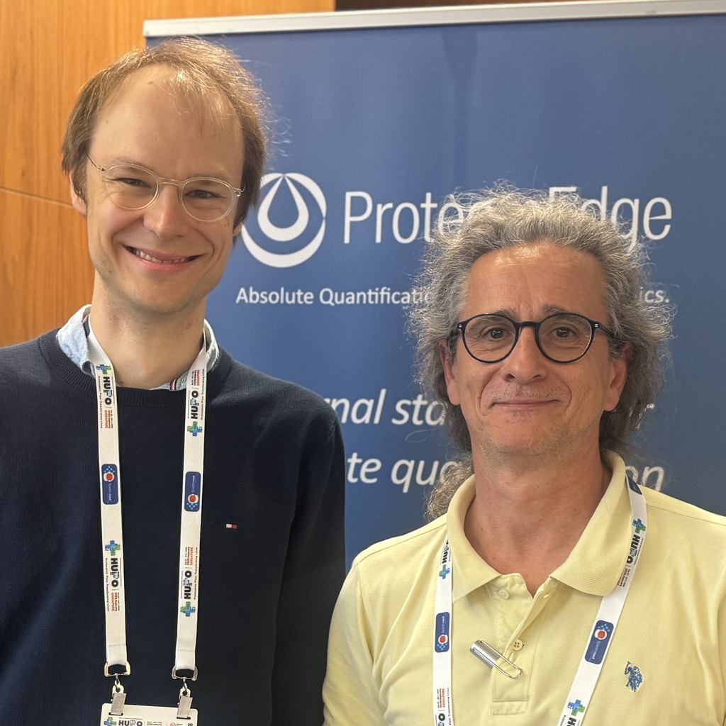
At the sensitivity limits of single-cell proteomics, small differences in sample handling and cell size can affect quantitative accuracy. Florian Rosenberger and Ákos Végvári of Karolinska Institutet discuss their evaluation of ProteomEdge qRePS™ at low sample loads, what they observed across two mass-spectrometry platforms, and why they want to bring internal standards into single-cell Deep Visual Proteomics. The interview was recorded at HUPO 2026 in Singapore and edited for length and clarity.
| Interviewees | Florian Rosenberger, Assistant Professor and group leader; Ákos Végvári, Senior Research Specialist in the Rosenberger group (Karolinska Institutet, Stockholm) |
| Stakeholder lens | Academic research and methods development in single-cell and spatial proteomics |
| ProteomEdge technology evaluated | ProteomEdge qRePS™ (Quantitative Recombinant Protein Standards); low-load evaluation using data-independent acquisition (DIA) |
| Platforms | Orbitrap Astral and timsTOF SCP |
| Public reference / use case | Interview recorded at HUPO 2026 in Singapore; evaluation of internal standards for low-load proteomics, with single-cell Deep Visual Proteomics as a proposed next step |
| Application area | Quantitative single-cell and spatial proteomics; future application to mitochondrial disease research |
Florian Rosenberger: In single-cell proteomics, we are operating the mass spectrometer at the lower end of its sensitivity. That makes us particularly vulnerable to everything that happens throughout the workflow. We might lose a little material during sample preparation, or have smaller or larger cells. These differences can result in non-linear effects that skew our quantitative accuracy.
By spiking in internal standards as early as possible in the workflow, we can control for these inaccuracies and obtain better quantitative data.
Ákos Végvári: We could detect the qRePS-derived peptides very well in data-independent acquisition mode, regardless of which mass spectrometer or vendor we used. The resulting peptides behaved very consistently.
We can also combine different sets of qRePS to target endogenous proteins. Adding protein standards early in the workflow creates the possibility of monitoring digestion efficiency, alongside more controlled and accurate quantification of endogenous peptides.
Our hope is that the reference peptides generated from qRePS will enable us to compare and normalise samples across different cell types and sample amounts.
Ákos Végvári: We learned that the standards were consistently detected across the mass-spectrometry platforms we tested. We used the same liquid chromatographic separation of the peptides, and both Orbitrap Astral and timsTOF SCP could sensitively detect low levels of qRePS-derived peptides.
We see this as a promising sign that these standards could be useful across different platforms for low-load and single-cell proteomics.
Florian Rosenberger: In Deep Visual Proteomics, we cut single cells from intact tissue specimens and measure them one at a time by mass spectrometry. This workflow is particularly sensitive to what happens throughout the process.
We might cut smaller or larger shapes, and protein retrieval may be better for some samples than for others. We are interested in bringing internal standards such as qRePS into our single-cell Deep Visual Proteomics workflow to help us trace that variation and improve quantitative accuracy at the end.
Florian Rosenberger: We think Deep Visual Proteomics and single-cell proteomics can be powerful in many disease settings. They are particularly useful where tissue is heterogeneous and the spatial arrangement of cells really matters.
Our focus is on mitochondrial diseases, where we often see tissue mosaics and every cell is its own individual. We want to understand how each cell responds to a particular defect, both in a failure mode and in a resilience mode. That is why we need to look at individual cells.